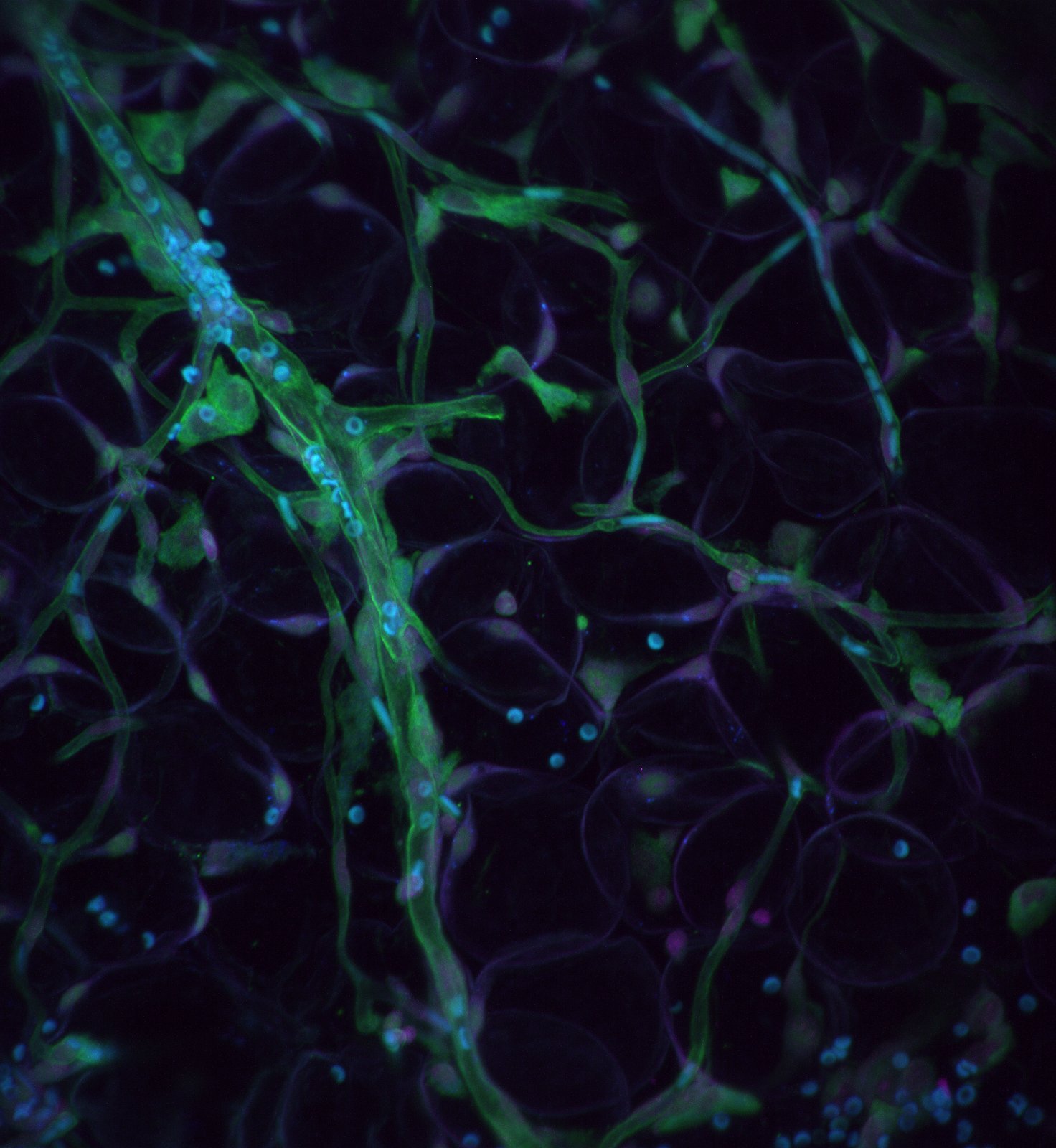
Fast, Gentle, Spectrally Multiplexed, 3D Imaging
Advanced Imaging Systems
At the UTSW-UNC Center for Cell Signaling Analysis, our team of researchers and engineers has set a clear and ambitious goal: to create and share affordable, high-performance 3D light-sheet microscopes. By combining cutting-edge technology with cost-effective solutions, we are dedicated to democratizing the field of microscopy and making it accessible to researchers worldwide. Through our collaborative efforts and commitment to innovation, we strive to empower scientists and accelerate scientific breakthroughs in cellular imaging and analysis.
Our approach involves…
Robust & performant open-source microscope control software.
Greatly simplified optoelectronic systems.
Highly engineered optical baseplates that enable easy assembly by non-experts.
Of paramount importance, our systems will be uniquely designed to harness the potential of advanced biosensors, chemogenetic and optogenetic modulators. Moreover, they will be readily compatible with state-of-the-art computer vision routines, affording unprecedented insights into the causal organization of biochemical signaling cascades in vivo. By empowering researchers to explore biological processes with heightened precision and analytical acumen, our microscopes hold the promise of transforming the landscape of scientific inquiry and discovery.
Microscope Technologies
-

Axially Swept Light-Sheet Microscopy
Imaging in physiological 3D extracellular matrix environments with an isotropic resolution of ~400 nm.
-

Field Synthesis Microscopy
Modular, reconfigurable light-sheet microscope that achieves lattice light-sheet microscopy performance in an easier-to-assemble & operate format.
-

Oblique Plane Microscopy
High-speed, large field of view microscopes that deliver the best lateral resolution in light-sheet microscopy.
navigate
To leverage advanced time-series analyses, deterministic microscope operation is paramount. To achieve this, we developed navigate, a comprehensive software package that is streamlined for non-expert use and includes key alignment and correction routines. Software interfaces seamlessly with the optogenetic and chemogenetic modules. All data is saved in cutting-edge Next-Generation File Formats with metadata according to the Open Microscopy Environment standard.
compass
COMPASS seeks to make high-resolution light-sheet fluorescence microscopy accessible to a wider audience. By integrating modular optomechanics, advanced lens simulations, and smart software, COMPASS facilitates the quick construction of light-sheet fluorescence microscopes that provide approximately 235 nm lateral and 465 nm axial resolution in an easy-to-use format.


