Visualizing, Controlling, and Quantifying Cellular Signaling

September 22 to 27, 2025

UT Southwestern Medical Center, Dallas, TX

After last year’s inaugural workshop for disseminating our tools and pipelines to trainers of microscopy cores, we are excited to announce our second workshop designed for advanced researchers—such as PhD students and postdocs—who are interested in integrating light-sheet microscopy, molecular imaging tools (biosensors, opto/ chemogenetics), and time-series analysis to quantitatively elucidate the intracellular signaling events that shape cell biological outcomes.

The six-day workshop will take place over consecutive days at UT Southwestern Medical Center in Dallas, TX. The first three days of the workshop will be structured as plenary sessions, where all participants engage in foundational training and interactive demonstrations. This will be followed by three days of small-group, hands-on activities, allowing participants to delve into one of three focus areas:

i) biosensors and opto / chemogenetic tools
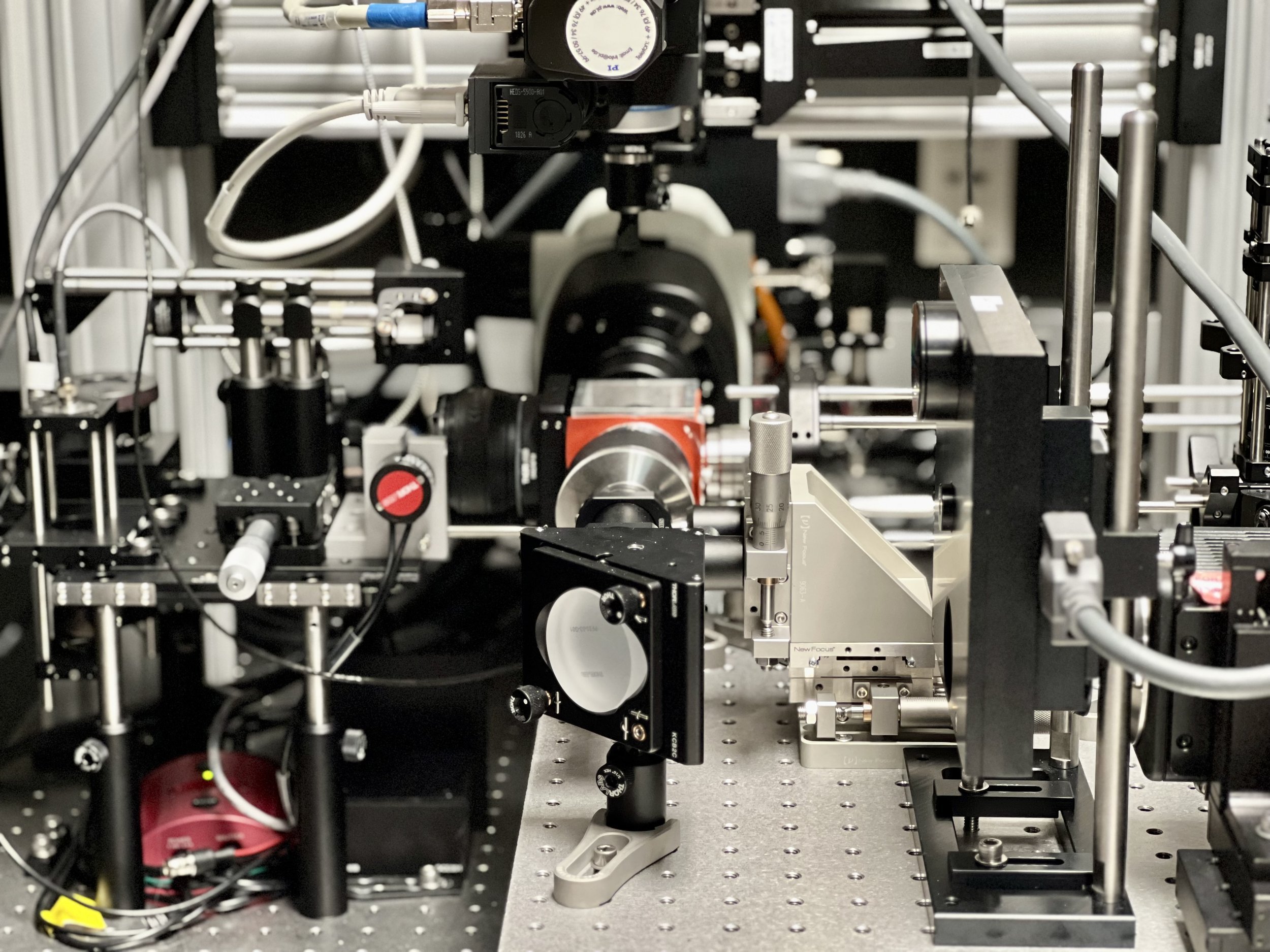
ii) light-sheet microscope construction
iii) live cell image analysis of signaling circuits using causal inference tools in a high-performance computing environment

Due to the hands-on nature of this workshop and to maintain a good ratio between instructors and participants for maximum interaction and personalized training, the capacity of our sessions are limited. Selection will be based on best fit between participant statements on the registration form and our workshop’s dissemination objectives.

Participation in the entire workshop is highly recommended, and applicants should be prepared to fully engage in both the large-group sessions and their chosen small-group focus.

Schedule

Focus
Sessions
Info

Application
Info

Other details

  • Learn about different types of probes and biosensors for tracking cell signaling events.

  • Learn about computational image analysis for cell signaling events.

  • Learn about the microscopes and smart imaging for cell signaling.

  • Choose to attend one of the three focus sessions depending upon your research and learning needs:
    Molecular Imaging Tools
    Computational Signaling Circuit Analysis
    Optics

Molecular Imaging Tools

The goal of the three days will be for participants to carry out their own experiments using different types of biosensors and chemo/optogenetic protein analogs. We will emphasize practical considerations and skills at the microscope, e.g. cell handling, responses of different cell types, irradiation with minimal perturbation, signal/noise etc. In parallel to this, participants can consult with center members about making tools to visualize or control protein activities of interest to them. Working with you, we will suggest designs for sensing, control, and multiplexed imaging of your proteins. For this, it would be helpful if you write to us in advance, letting us know what you are trying to accomplish and when possible including structure and function information.

Computational Signaling Circuit Analysis

The goal of the three days is to provide participants with insights of the theory behind the algorithms and hands-on experience with various packages of the u-analyze software suite. Emphasis will be put on methods for high-precision cell segmentation in 2D and 3D, extraction of subcellular time-series in shape- and motion-agnostic coordinate systems, and the inference of correlative and causal relations between signaling events and cellular behaviors in order to reconstruct control circuits of cell regulation. We will also provide instructions on the architecture of u-analyze to empower participants in extending the software for their own needs.

Optics

The goal of this three-day session is to provide participants with hands-on experience in building and optimizing a complete light-sheet microscopy system for live-cell imaging. Participants will configure all optics, optoelectronics, and optomechanics, gaining practical skills in assembling, aligning, and calibrating advanced imaging systems. Users will also learn how to use our open-source software, navigate, to control the microscope and automate imaging. Additionally, routine image post-processing steps, including deconvolution and data visualization, will be covered. Participants will have the opportunity to consult with CCSA staff to refine their own imaging setups and discuss strategies for optimizing microscope performance. If you have specific optical challenges or design considerations in your research, we encourage you to contact us in advance to tailor the workshop experience to your needs.

Applications will be accepted until 5/15/2025. Decisions regarding admission will be sent out by 6/2/2025.
Due to the hands-on nature of this workshop and to maximize participant learning, our plenary sessions are capped at 20 and each focus session at 6 participants.
Please note that you’ll need to provide your research question along with any information you may wish to communicate regarding probes of interest, considerations for imaging, sample types, etc. for the focus sessions. You will be able to select only one of the three focus sessions.

Participants are expected to fund their own travel to Dallas and lodging. Upon acceptance, we will distribute a list of stay options near UT Southwestern.

Lunch (and dinner on longer days) is covered. There are no fees to attend; just the above mentioned self-funded travel and lodging.

Questions? Email us.